Complex plasmon-exciton dynamics revealed through quantum dot light emission in a nanocavity

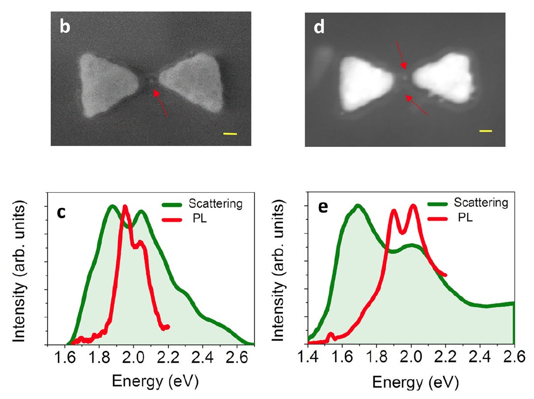
Plasmonic cavities can confine electromagnetic radiation to deep sub-wavelength regimes. This facilitates strong coupling phenomena to be observed at the limit of individual quantum emitters. Here, we report an extensive set of measurements of plasmonic cavities hosting one to a few semiconductor quantum dots. Scattering spectra show Rabi splitting, demonstrating that these devices are close to the strong coupling regime. Using Hanbury Brown and Twiss interferometry, we observe non-classical emission, allowing us to directly determine the number of emitters in each device. Surprising features in photoluminescence spectra point to the contribution of multiple excited states. Using model simulations based on an extended Jaynes-Cummings Hamiltonian, we find that the involvement of a dark state of the quantum dots explains the experimental findings. The coupling of quantum emitters to plasmonic cavities thus exposes complex relaxation pathways and emerges as an unconventional means to control dynamics of quantum states.

Authors:

Satyendra Nath Gupta, Ora Bitton, Tomas Neuman, Ruben Esteban, Lev Chuntonov, Javier Aizpurua & Gilad Haran

Read More

Glutathione Self-Assembles into a Shell of Hydrogen-Bonded Intermolecular Aggregates on “Naked” Silver Nanoparticles

A detailed understanding of the molecular structure in nanoparticle ligand capping layers is crucial for their efficient incorporation into modern scientific and technological applications. Peptide ligands render the nanoparticles as biocompatible materials. Glutathione, a γ-ECG tripeptide, self-assembles into aggregates on the surface of ligand-free silver nanoparticles through intermolecular hydrogen bonding and forms a few nanometer-thick shells. Two-dimensional nonlinear infrared (2DIR) spectroscopy suggests that aggregates adopt a conformation resembling the β-sheet secondary structure. The shell thickness was evaluated with localized surface plasmon resonance spectroscopy and X-ray photoelectron spectroscopy. The amount of glutathione on the surface was obtained with spectrophotometry of a thiol-reactive probe. Our results suggest that the shell consists of ∼15 stacked molecular layers. These values correspond to the inter-sheet distances, which are significantly shorter than those in amyloid fibrils with relatively bulky side chains, but are comparable to glycine-rich silk fibrils, where the side chains are compact. The tight packing of the glutathione layers can be facilitated by hydrogen-bonded carboxylic acid dimers of glycine and the intermolecular salt bridges between the zwitterionic γ-glutamyl groups. The structure of the glutathione aggregates was studied by 2DIR spectroscopy of the amide-I vibrational modes using 13C isotope labeling of the cysteine carbonyl. Isotope dilution experiments revealed the coupling of modes forming vibrational excitons along the cysteine chain. The coupling along the γ-glutamyl exciton chain was estimated from these values. The obtained coupling strengths are slightly lower than those of native β-sheets, yet they appear large enough to point onto an ordered conformation of the peptides within the aggregate. Analysis of the excitons’ anharmonicities and the strength of the transition dipole moments generally is in agreement with these observations.

Authors: Arghyadeep Basu, Alexander Vaskevich, and Lev Chuntonov*

Read More